Why HigHNoon?
In interventional radiology, every millimeter matters. Even a small deviation in needle angle or entry point can require additional scans, increasing radiation exposure and patient discomfort.
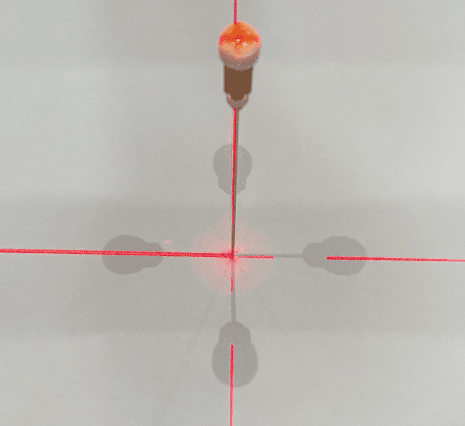
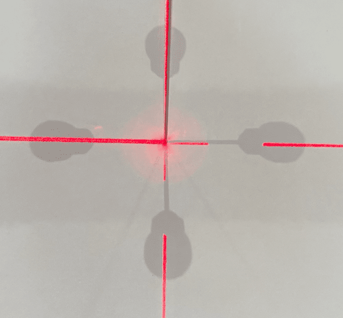
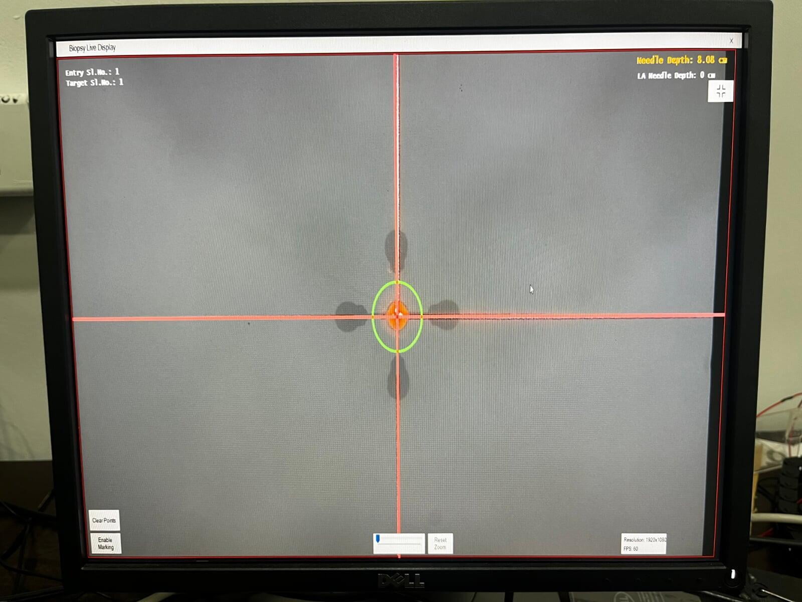
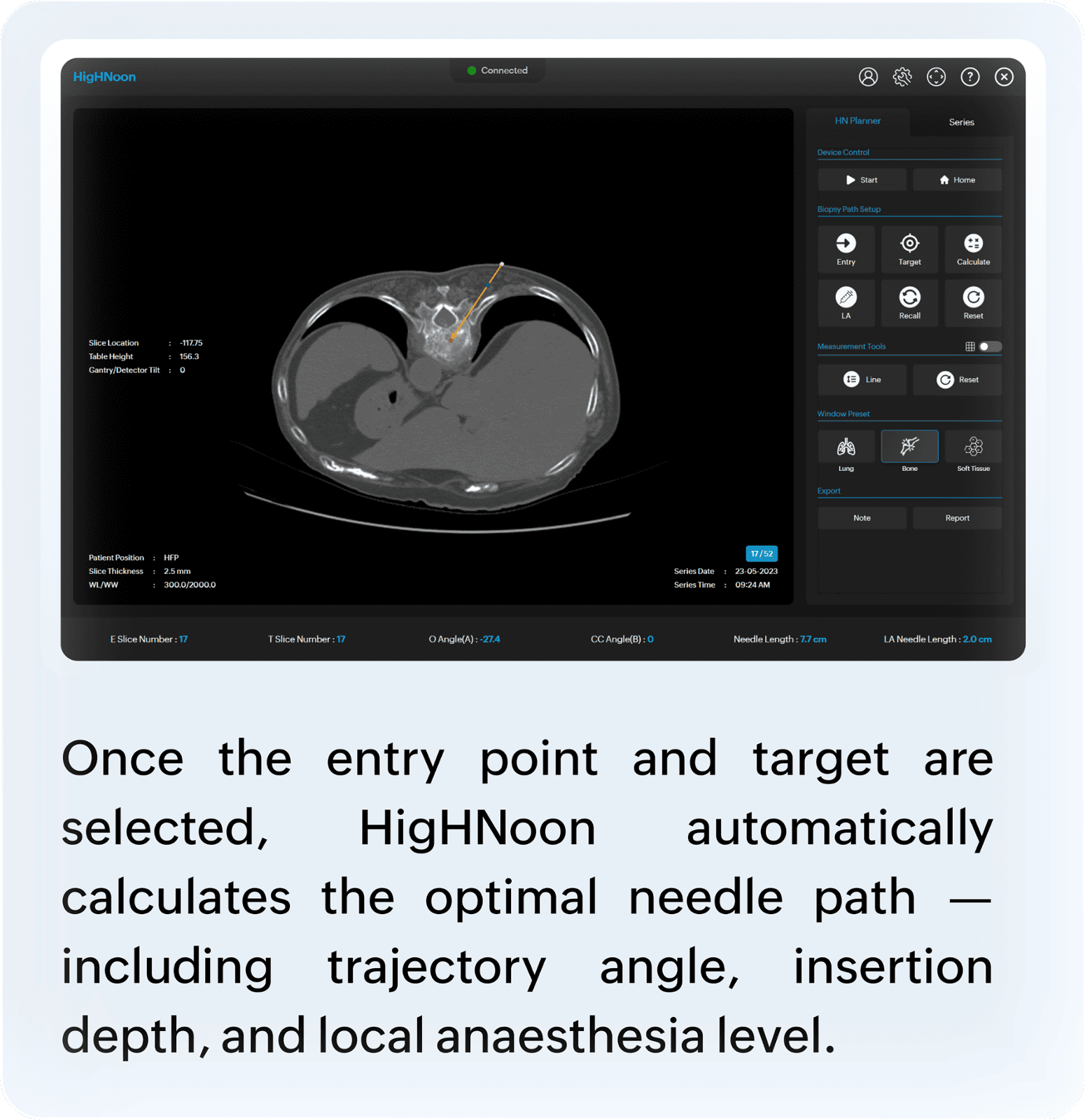
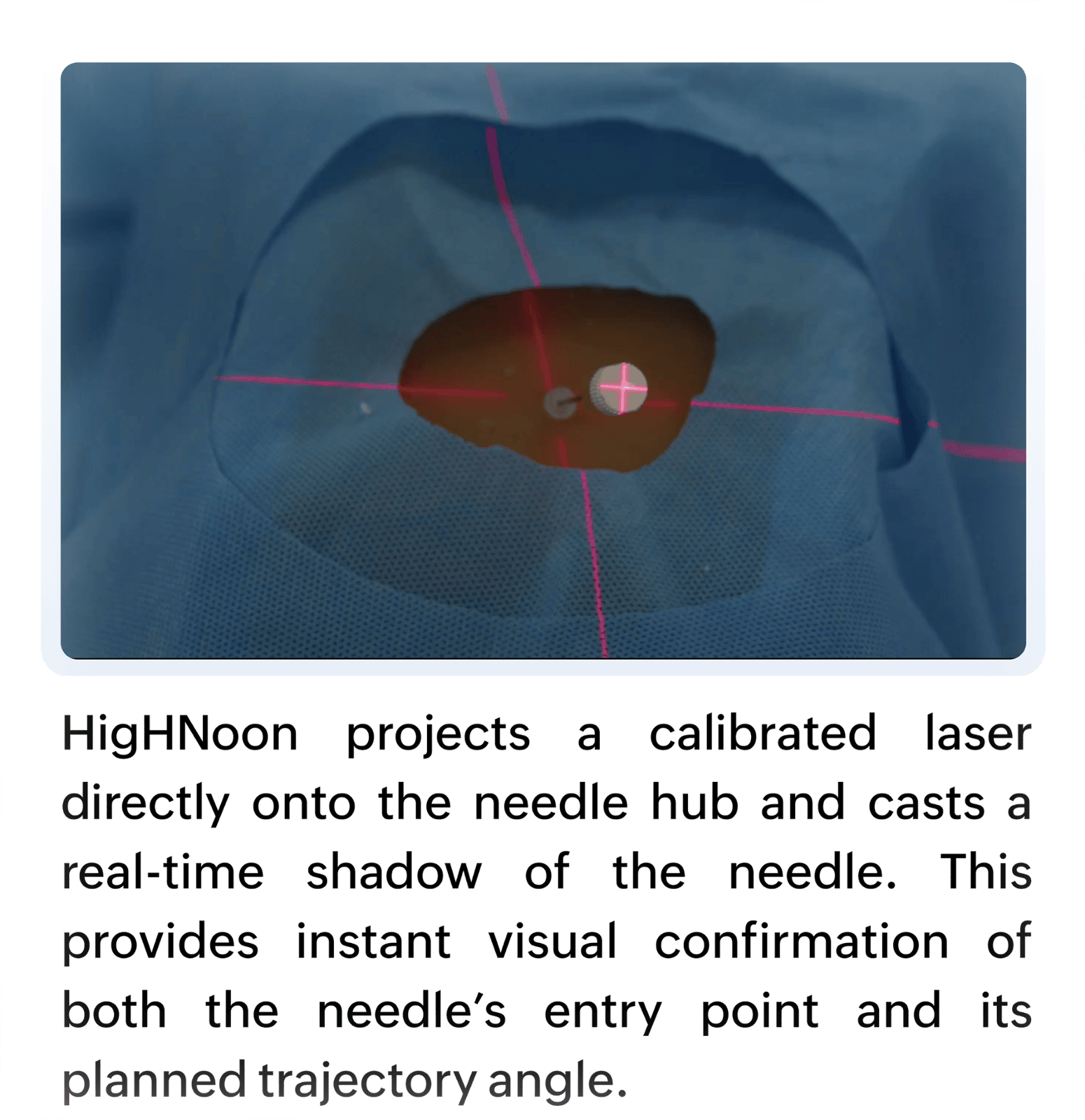
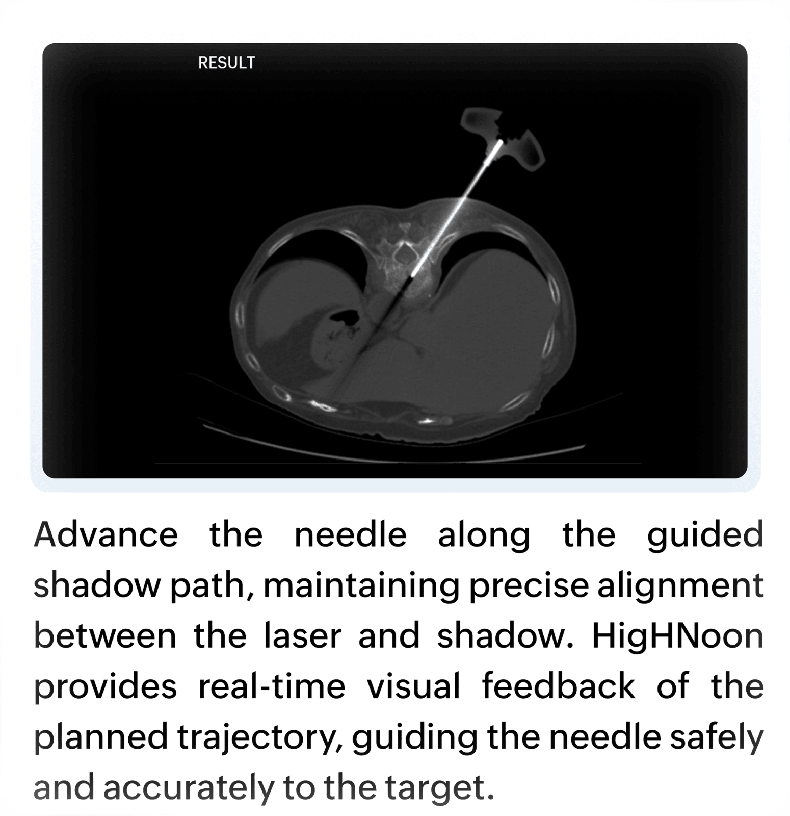
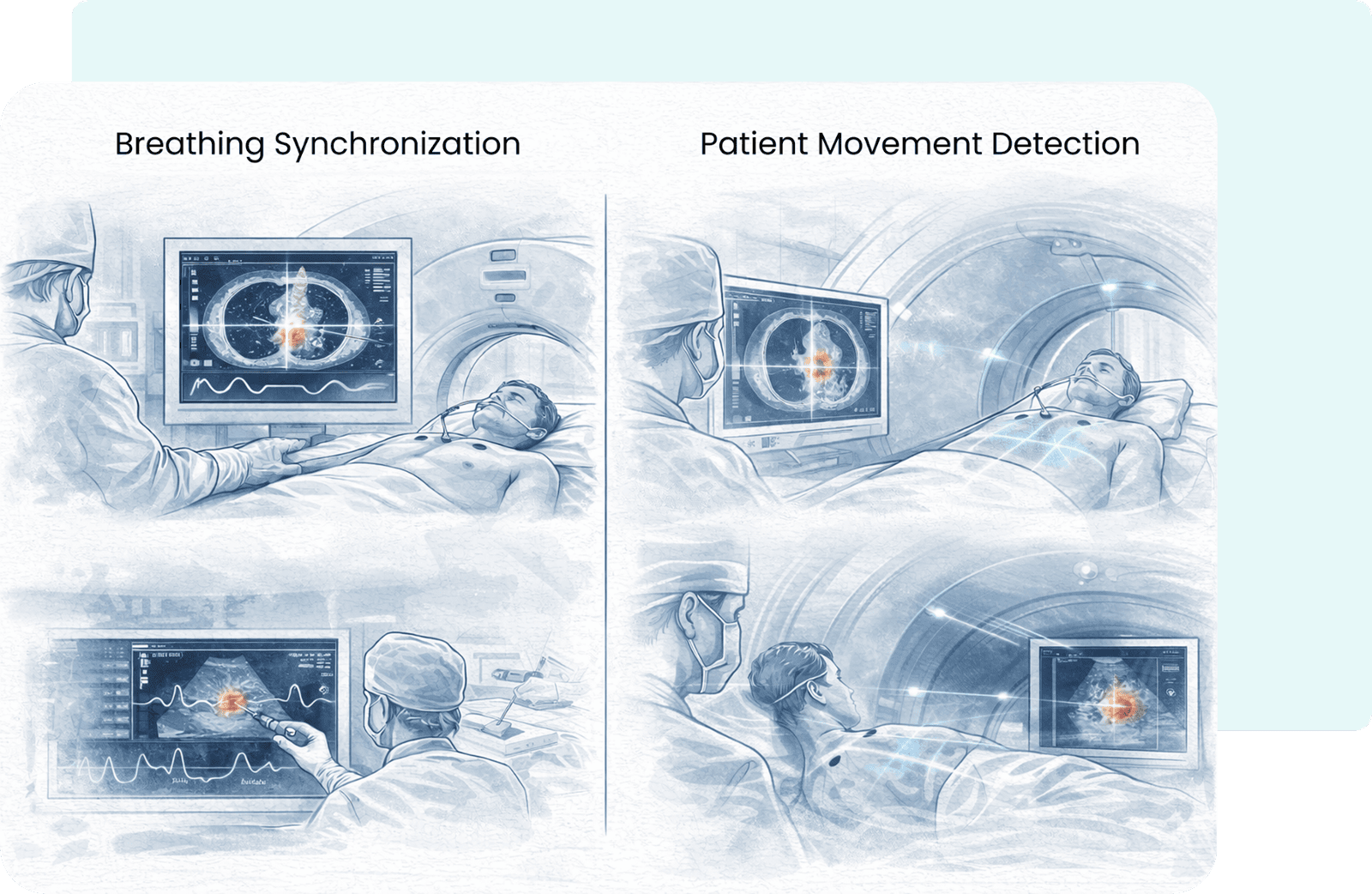
HigHNoon changes that. It transforms CT-guided interventions using light- and shadow-based real-time needle navigation. A calibrated light and laser system projects a precise shadow and laser guidance pattern aligned with the planned needle trajectory. As the clinician positions the needle, the shadow provides instant visual feedback, ensuring accurate alignment with the planned path before puncture and during insertion.
Real-Time Needle Guidance
High Positional Accuracy
Reduced Needle Manipulations
Shorter Procedure Time
Lower Radiation Exposure
Universal Compatibility
Non-Invasive Guidance
Check out HighNoon
Effortless Integration into Your Existing Workflow
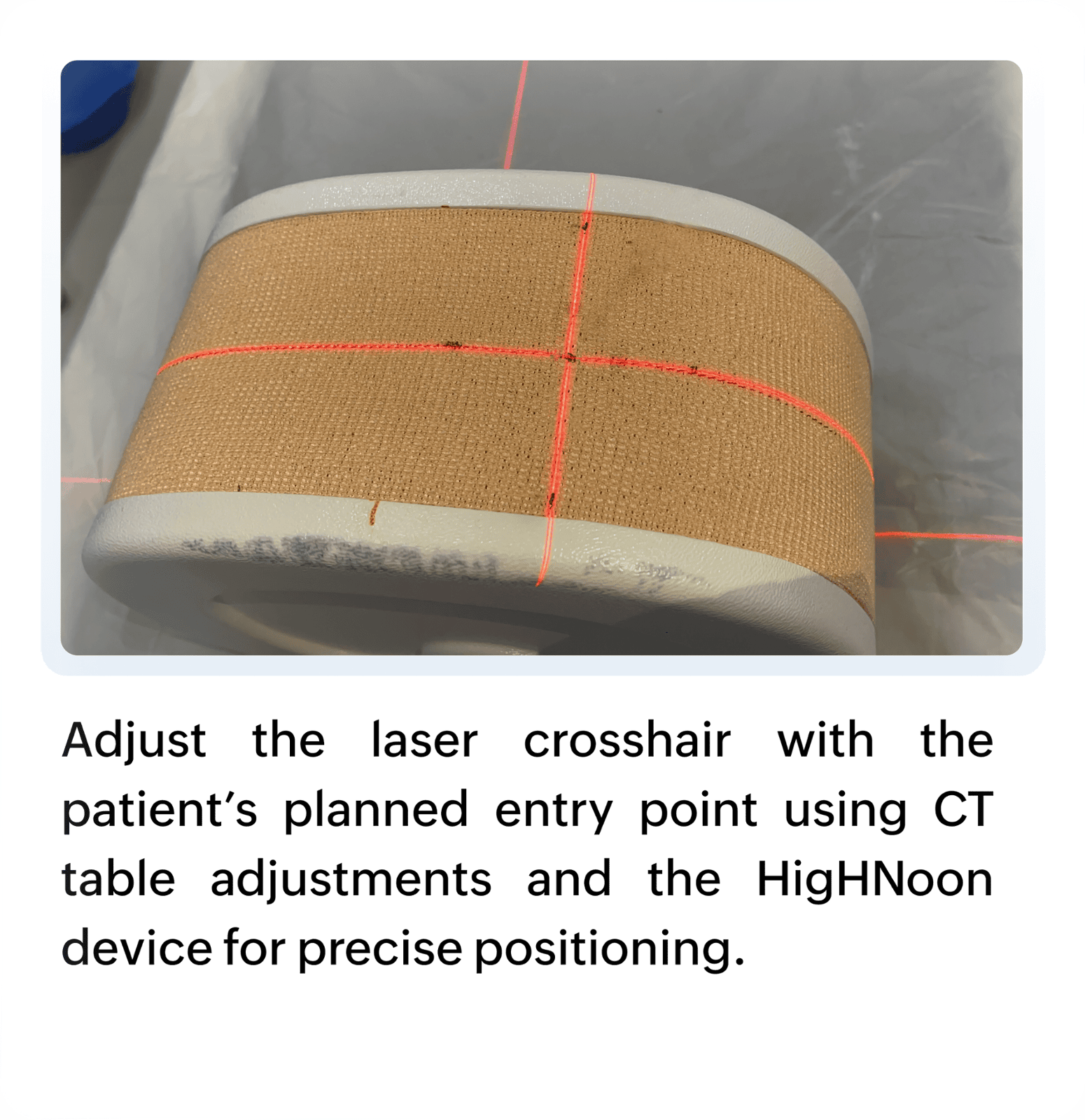
HigHNoon fits seamlessly into your CT-guided procedure — aligning with your existing planning and puncture steps.
Plan
Align
Guidance
Procedure
Three Key Pillars
Absolute Precision
Experience 2mm accuracy with real-time laser and shadow guidance. HigHNoon visually aligns every planned trajectory, ensuring the needle follows the exact CT-defined path — even in small, deep, or mobile targets.
Consistent Control
HigHNoon provides real-time visual guidance that keeps the planned trajectory aligned from entry to target—reducing variability and improving reproducibility. With fewer manipulations and rescans, procedures become faster, safer, and more consistent, while still preserving freehand flexibility.
Seamless Efficiency
HigHNoon integrates seamlessly with any existing CT workflows. Its plug-and-play design requires no repeated calibration, enabling quick setup and supporting multiple clinical applications without disrupting routine operations.
Clinical Benefits:
For interventional radiologists, HigHNoon simplifies complex needle-guided procedures and enhances operator confidence—even for deep-seated, small, or critically located targets.
For patients, it means:
- Shorter time on the CT table
- Less radiation exposure
- Fewer punctures
- Higher accuracy and comfort
Patient-Centric Features
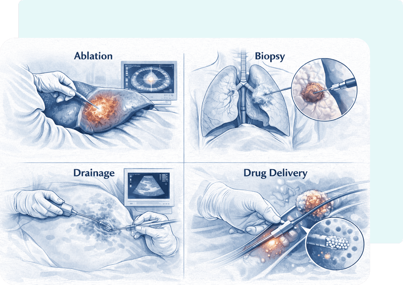
Clinical Applications:
- Ablation
- Biopsy
- Drainage
- Drug Delivery
Clinical Experience:
HigHNoon has shown exceptional performance in a wide range of CT-guided interventions — from complex biopsies to targeted ablations.
Its real-time laser and shadow guidance ensures precise, repeatable accuracy across all organs and anatomies. This reduces rescans, minimizes radiation exposure, and lowers procedural variability, helping radiologists perform safer and more efficient interventions.
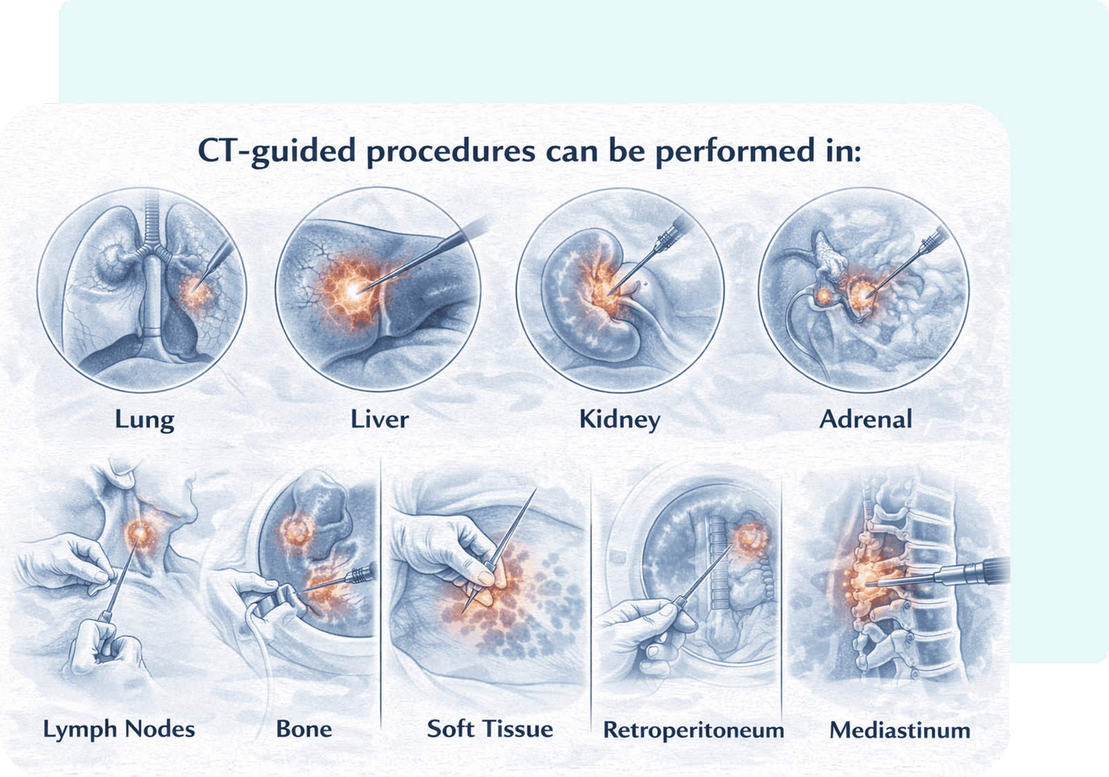
CT-guided procedures can be performed in:
•Lung • Liver • Kidney • Adrenal • Lymph Nodes • Bone • Soft Tissue • Retroperitoneum • Pelvis • Mediastinum • SpineClinical Outcomes
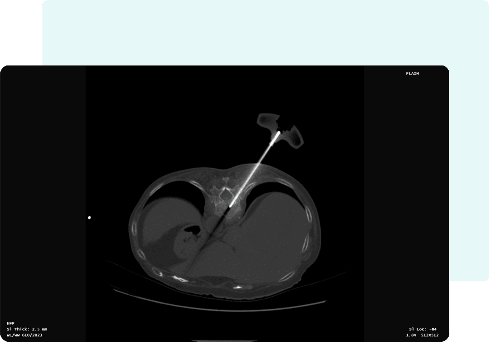
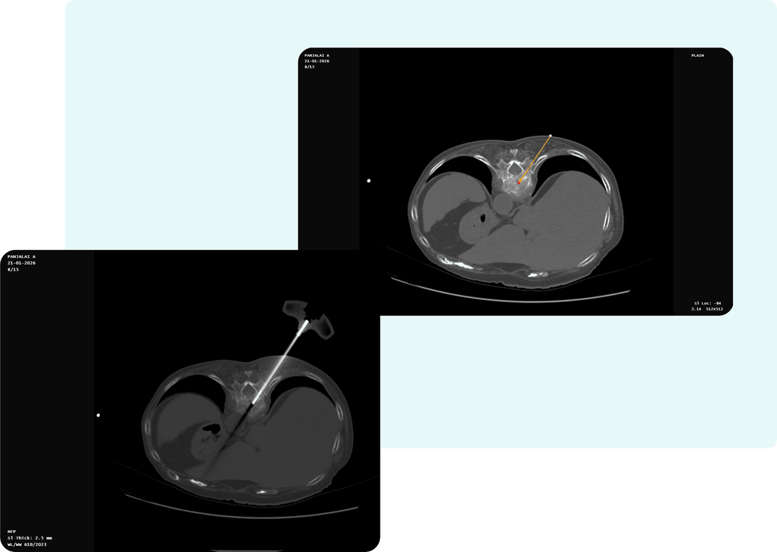
- Needle Length: 6.65 cm
- Entry Slice Number: 8
- Target Slice Number: 8
- Orbital Angle: 33.99°
- Cranio-Caudal (CC) Angle: 0°
Clinical Outcomes
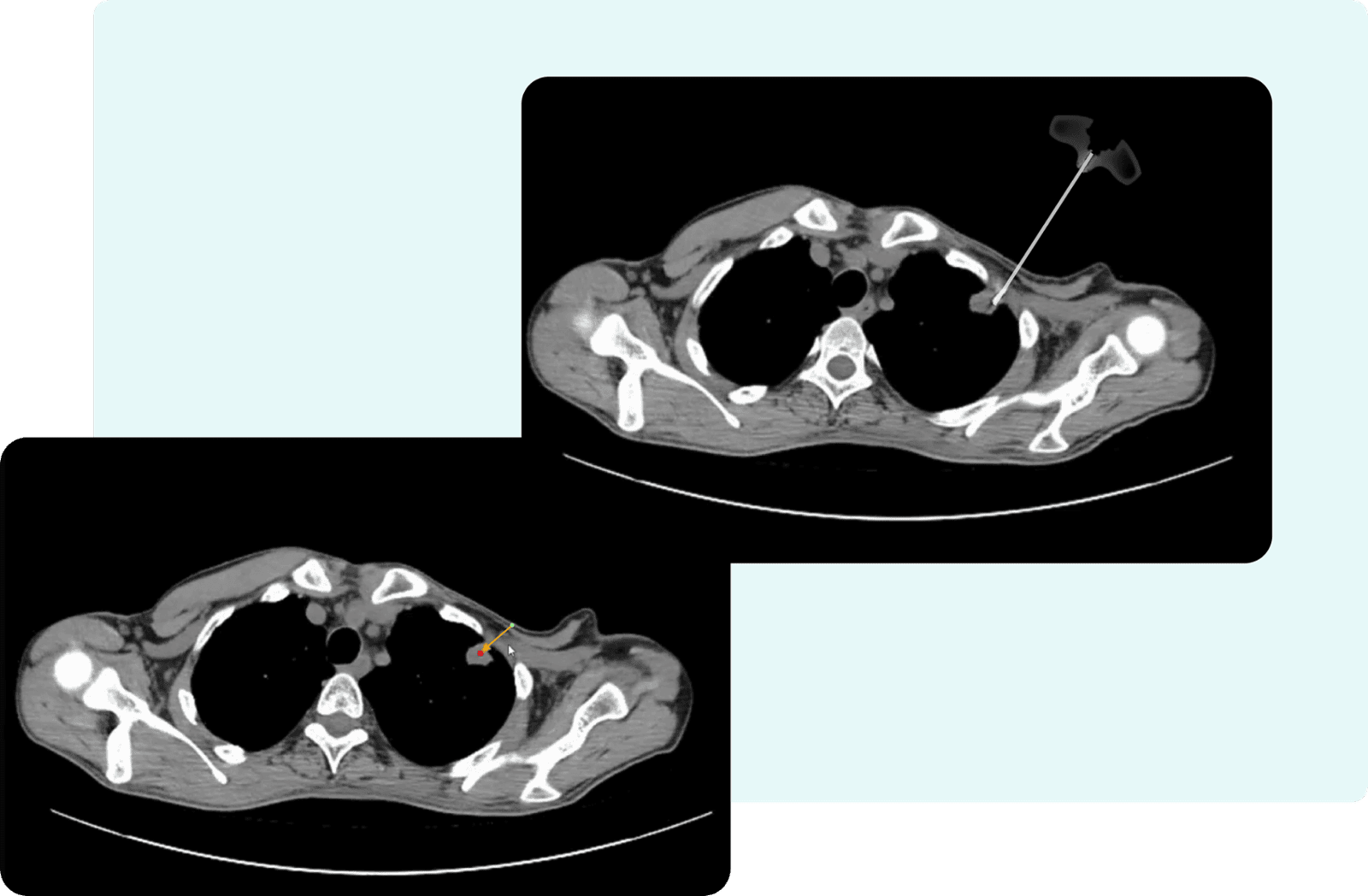
- Needle Length: 2.03cm
- Entry Slice Number: 8
- Target Slice Number: 10
- Orbital Angle: 42.27°
- Cranio-Caudal (CC) Angle: -18.93°
Publication
Efficacy of Shadow-Based Needle Positioning System in Performing CT Image-Guided Percutaneous Biopsy of Lung Lesions: Our Initial Experience
Computed tomography (CT) plays a key role in interventions such as tumor ablation, FNAC/FNAB, percutaneous block of the upper sympathetic chain, treatment of secondary pulmonary aspergilloma, and brachytherapy. The procedures are user-dependent, and achieving accuracy is challenging. Several navigation tools have been innovated for CT-guided interventions; however, drawbacks continue to exist. There is a need for a navigation tool with improved accuracy and reduced intervention time.